Blood Vessels Labeled : Pin On Nursing : The thick outermost layer of a vessel (tunica adventitia or tunica externa) is made of connective tissue.. Blood vessels labeled diagram : The major arteries in the body. Arteries, arterioles, capillaries, venules and veins. Arteries carry blood away from the heart to other organs. Name the blood vessel labeled 'c'.
A vein is a blood vessel that conducts blood toward the heart. Arteries and veins are composed of three tissue layers. •formed where capillaries unite • extremely porous 1) venules: Name the blood vessels labeled 'e'. Between arteries and veins, there is a network of.
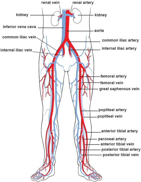
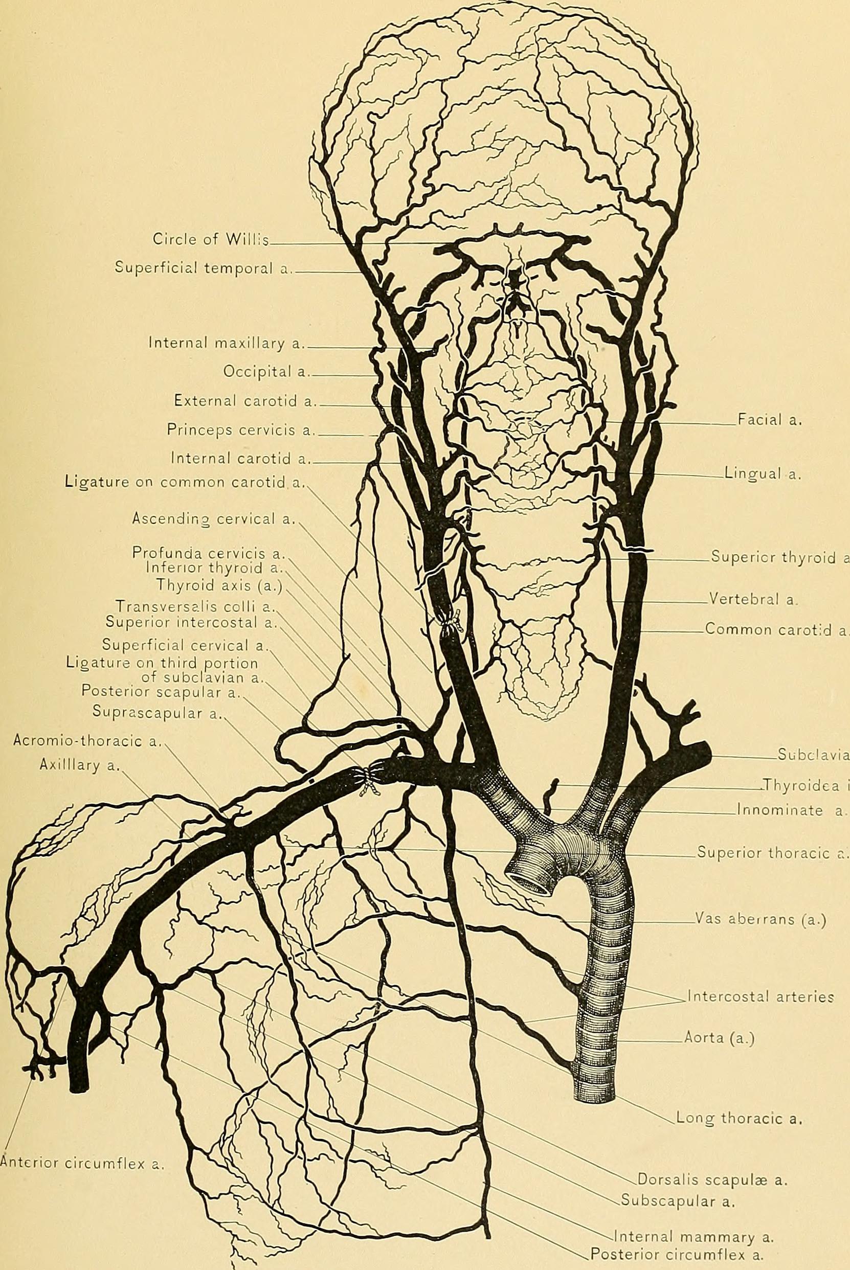
Labeled Picture Of The Nervous System Labeled Nervous System Labeled Diagram Nervous System Human Diagram Koibana Info Nervous System Anatomy Medical Anatomy Human Body Anatomy from i.pinimg.com The superior vena cava is the large vein that brings blood from the head and arms to the heart, and the inferior vena cava brings blood from the abdomen and legs into the heart. Figures 1 and 2 show the major arteries and veins of the body. Blood vessels consist of arteries, arterioles, capillaries, venules, and veins. There are five main types of blood vessels: Between arteries and veins, there is a network of. Very small branches that collect the blood from the various organs and parts are called venules, and they unite to form veins, which return the blood to the heart. Bulky middle tunic contains smooth muscle and elastin 3. Blood is supplied to parts within the neck, head and brain through branches of the subclavian and common carotid arteries.
The vessels that carry blood away from the heart are called arteries, and their very small branches are arterioles.
Arteries (in red) are the blood vessels that deliver blood to the body. Blood is supplied to the brain, face, and scalp via two major sets of vessels: Blood vessels labeled diagram : Select this option to access the mini anatomy model of the blood vessels. Normal function of the brain's control centers is dependent upon adequate supply of oxygen and nutrients through a dense network of blood vessels. Blood vessels are found throughout the body. Hma practical 3 for monday july 23 and wednesday july 25. The inner lining is the endothelium and is surrounded by subendothelial connective tissue. The aorta is the largest and closest to the heart, beginning right after the aortic valve. A demonstration of the major arteries and veins of the human body for human anatomy and physiology Eventually, the smallest arteries, vessels called arterioles, further branch into tiny capillaries, where nutrients and wastes are exchanged, and then combine with other vessels that exit capillaries to form venules, small blood vessels that carry blood to a vein, a larger blood vessel that returns blood to the heart. The major arteries in the body. Between arteries and veins, there is a network of.
The major veins in the Label the arteries of the aortic arch in the ct angiogram. Hma practical 3 for monday july 23 and wednesday july 25. Use key choices to identify the blood vessel tunic described. A vein is a blood vessel that conducts blood toward the heart.
Illustrations Of The Blood Vessels from my.clevelandclinic.org The right and left common carotid arteries and the right and left vertebral arteries. This set is often in folders with. Label the blood vessels and structures using the hints provided. The major veins in the The thick outermost layer of a vessel (tunica adventitia or tunica externa) is made of connective tissue. Anatomy of blood vessels review sheet 32 261 microscopic structure of the blood vessels 1. Label the arteries of the aortic arch in the ct angiogram. Arteries and veins are composed of three tissue layers.
Select this option to access the mini anatomy model of the blood vessels.
There are five main types of blood vessels: Blood is supplied to the brain, face, and scalp via two major sets of vessels: This set is often in folders with. A demonstration of the major arteries and veins of the human body for human anatomy and physiology The function and structure of each segment of the peripheral vascular system vary depending on the organ it supplies. Arteries carry blood away from the heart to other organs. Arteries, arterioles, capillaries, venules and veins. This article lists a series of labeled imaging anatomy cases by system and modality. Related posts of the human blood vessels labeled digestive system orangs with function. Blood circulates throughout the body in blood vessels, propelled by the pumping action of the heart. Label the arteries of the aortic arch in the ct angiogram. •formed where capillaries unite • extremely porous 1) venules: Digestive system orangs with function 12 photos of the digestive system orangs with function digestive system and organs with function, digestive system organs and functions ppt, digestive system organs and functions quiz, digestive system organs and functions table, digestive system with organs and.
Aside from capillaries, blood vessels are all made of three layers: Blood vessels are vital for the body and play a key role in diabetes helping to transport glucose and insulin. The superior vena cava is the large vein that brings blood from the head and arms to the heart, and the inferior vena cava brings blood from the abdomen and legs into the heart. The common cartoid artery extends from the brachiocephalic artery. The vessels that carry blood away from the heart are called arteries, and their very small branches are arterioles.
Blood Vessels 3 Labeled Blood Vessels 3 Labeled Brachial Vein Basilic Vein Cephalic Vein Median Cubital V Accessory Cephalic V Cephalic Vein Sup Course Hero from www.coursehero.com Label the arteries of the aortic arch in the ct angiogram. Figures 1 and 2 show the major arteries and veins of the body. The venules and veins returning blood to the heart. Use key choices to identify the blood vessel tunic described. The thick outermost layer of a vessel (tunica adventitia or tunica externa) is made of connective tissue. Select this option to access the mini anatomy model of the blood vessels. Access the model when a vein or artery is selected, access to the detailed view of the blood vessels is available. The vessels allow blood to be pumped at a high pressure to deliver nutrients and.
Name the blood vessel labeled 'd'.
Capillaries are blood vessels that are one cell thick (endothelium) where the main diffusion and exchange. They are designated as resistance vessels since they can regulate blood flow velocity by means of their respective muscle walls (approximately 120 mm hg). Blood vessels and lymph nodes. This set is often in folders with. The common cartoid artery extends from the brachiocephalic artery. The aorta is the largest and closest to the heart, beginning right after the aortic valve. The 4 valves are the aortic, pulmonary, mitral, and tricuspid valves. There are three main types of blood vessels:. It extends on each side of the neck and divides at the level of the larynx into two branches: Blood circulates throughout the body in blood vessels, propelled by the pumping action of the heart. To play this quiz, please finish editing it. There are five main types of blood vessels: Access the model when a vein or artery is selected, access to the detailed view of the blood vessels is available.
How Many Carotid Arteries In The Neck : Department Of Surgery Carotid Artery Disease - In anatomy, the left and right common carotid arteries (carotids) (english:. Carotid artery, one of several arteries that supply blood to the head and neck. Effect of patching on reducing restenosis in the carotid revascularization endarterectomy versus stenting trial. As a very rare occurence, the carotid my ascend in the neck without dividing into the two usual branches; The internal carotid artery is a major branch of the common carotid artery, supplying several parts of the head with blood, the most important one being the brain. Cervical arterial dysfunction , carotid arteries, manipulation, manual therapy, neck, stroke, vascular.
The left arises directly from the aorta, a large artery arising from the heart. The internal carotid artery (latin: The cervical part of the internal carotid artery undergoes a straight course in the neck from its origin up to the base of skull. How do we come to know that our artery is damaged? Vertebral arteries (arteria vertebralis) next to your spine which become basilar artery (arteria.
Arteries In The Neck The Carotid Arterial System Lecturio from d3uigcfkiiww0g.cloudfront.net In the neck, each of them branches off into an internal carotid artery. They ascend in the neck the left common carotid varies in its origin more than the right. Its origin is more in the internal part of the. The common carotid artery sometimes follows a very tortuous course, forming one or more distinct loops in the neck. Either the external or internal carotid being absent. How many carotid endarterectomies are performed each year? The carotid sinus contains sensors that help regulate blood pressure. When a person has a stroke it can be from blockage in 1 or both of the carotid arteries in the neck.
There are arteries in the neck as shown in the image.
There are two internal carotid arteries in total, one on each side of the neck. Carotid artery stenosis, known more commonly as carotid artery disease, is a result of the narrowing or blocking of the arteries that leads to a decrease the blood clots and deposits can become trapped in smaller blood vessels in the brain, decreasing blood supply to the area. Many anatomical features add risk to surgery, and these include previous neck irradiation, a contralateral carotid occlusion or efficacy of embolic protection devices: The left arises directly from the aorta, a large artery arising from the heart. When a person has a stroke it can be from blockage in 1 or both of the carotid arteries in the neck. There are arteries in the neck as shown in the image. A blockage in one of the carotid arteries can be cleared either by endarterectomy or carotid angioplasty. Therapeutic applications in the management of advanced. Branch of in the thoracic section, the left common carotid travels upwards through the superior mediastinum to the level of the left sternoclavicular joint where it. Important risk factors in addition to the degree of stenosis include, gender, diabetes, the type of stroke symptoms (whether in the brain or in the eye), blockage of the carotid artery on the opposite side, and the use of aspirin immediately. Vertebral arteries (arteria vertebralis) next to your spine which become basilar artery (arteria. They can be called the main arteries of the head and neck. The principal arteries of supply to the head and neck are the two common carotids;
Vertebral arteries (arteria vertebralis) next to your spine which become basilar artery (arteria. Effect of patching on reducing restenosis in the carotid revascularization endarterectomy versus stenting trial. Moving from the sternoclavicular joint, both sides move upwards along a slanting path to the upper border of the thyroid cartilage in the neck. The principal arteries of supply to the head and neck are the two common carotids; It supplies structures present in the cranial cavity and orbit.
Carotid Triangle Anatomy Kenhub from thumbor.kenhub.com Effect of patching on reducing restenosis in the carotid revascularization endarterectomy versus stenting trial. The common carotid artery sometimes follows a very tortuous course, forming one or more distinct loops in the neck. The internal carotid artery is a major branch of the common carotid artery, supplying several parts of the head with blood, the most important one being the brain. Origin the right common carotid artery originates behind the sternoclavicular. The cervical part of the internal carotid artery undergoes a straight course in the neck from its origin up to the base of skull. Carotid artery stenosis, known more commonly as carotid artery disease, is a result of the narrowing or blocking of the arteries that leads to a decrease the blood clots and deposits can become trapped in smaller blood vessels in the brain, decreasing blood supply to the area. In the neck and head exterior to the skull, the external carotid artery provides blood flow to the skin, muscles, and organs. The carotid arteries are a pair of large blood vessels that carry blood from the heart to the brain.
The principal arteries of supply to the head and neck are the two common carotids;
They originate from the carotid bifurcation, travel through the. Vertebral arteries (arteria vertebralis) next to your spine which become basilar artery (arteria. When a person has a stroke it can be from blockage in 1 or both of the carotid arteries in the neck. Many anatomical features add risk to surgery, and these include previous neck irradiation, a contralateral carotid occlusion or efficacy of embolic protection devices: Carotid artery, one of several arteries that supply blood to the head and neck. The carotid artery pulse can normally be felt in the neck by pressing the fingertips against the side of the windpipe, or trachea. The right and left common carotid arteries have differing origins. Effect of patching on reducing restenosis in the carotid revascularization endarterectomy versus stenting trial. The right originates in the brachiocephalic trunk, the largest branch. As a very rare occurence, the carotid my ascend in the neck without dividing into the two usual branches; Carotid artery stenosis, known more commonly as carotid artery disease, is a result of the narrowing or blocking of the arteries that leads to a decrease the blood clots and deposits can become trapped in smaller blood vessels in the brain, decreasing blood supply to the area. Therapeutic applications in the management of advanced. The left arises directly from the aorta, a large artery arising from the heart.
Therapeutic applications in the management of advanced. Divisions of the carotid artery. When a person has a stroke it can be from blockage in 1 or both of the carotid arteries in the neck. After making an incision along the front of your neck, the surgeon opens the malas m, et al. In the neck and head exterior to the skull, the external carotid artery provides blood flow to the skin, muscles, and organs.
Internal Carotid Artery Anatomy Segments And Branches Kenhub from thumbor.kenhub.com Arteria carotis interna) is located in the inner side of the neck in contrast to the external carotid artery. They can be called the main arteries of the head and neck. How many carotid arteries are there? The internal carotid artery (latin: After making an incision along the front of your neck, the surgeon opens the malas m, et al. The common carotid artery sometimes follows a very tortuous course, forming one or more distinct loops in the neck. Vertebral arteries (arteria vertebralis) next to your spine which become basilar artery (arteria. The carotid sinus contains sensors that help regulate blood pressure.
Origin the right common carotid artery originates behind the sternoclavicular.
They can be called the main arteries of the head and neck. Both common carotid arteries run within the carotid sheath until they bifurcate into the internal and external carotid arteries at the level of the upper intraoperative radiation of canine carotid artery, internal jugular vein, and vagus nerve. When an area of the brain. It ascends up in the. There are two internal carotid arteries in total, one on each side of the neck. Either the external or internal carotid being absent. The right and left carotid arteries are the related questions. How many carotid endarterectomies are performed each year? It supplies structures present in the cranial cavity and orbit. The carotid artery pulse can normally be felt in the neck by pressing the fingertips against the side of the windpipe, or trachea. /kəˈrɒtɪd/) are arteries that supply the head and neck with oxygenated blood; The carotid arteries are a pair of large blood vessels that carry blood from the heart to the brain. The main arteries in the neck are the common carotids, and the main veins of the neck that return the blood scalenus posterior, the smallest and most deeply seated of the 3 scaleni, arises, by 2 or 3 separate tendons, from the posterior tubercles of the transverse processes of the lower 2 or 3 cervical.