Blood Vessels Labeled : Pin On Nursing : The thick outermost layer of a vessel (tunica adventitia or tunica externa) is made of connective tissue.. Blood vessels labeled diagram : The major arteries in the body. Arteries, arterioles, capillaries, venules and veins. Arteries carry blood away from the heart to other organs. Name the blood vessel labeled 'c'.
A vein is a blood vessel that conducts blood toward the heart. Arteries and veins are composed of three tissue layers. •formed where capillaries unite • extremely porous 1) venules: Name the blood vessels labeled 'e'. Between arteries and veins, there is a network of.
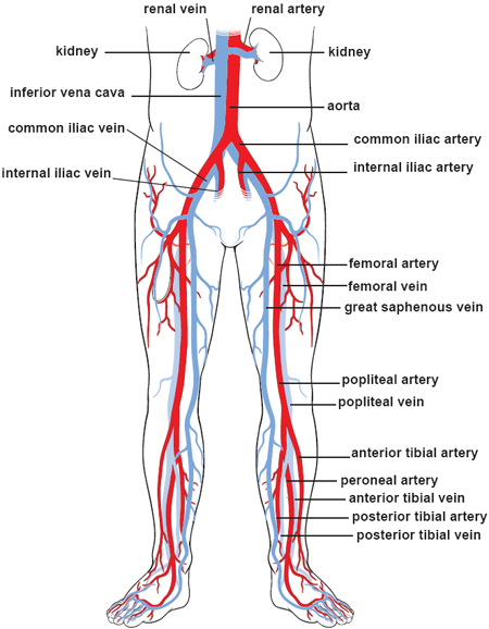
Labeled Picture Of The Nervous System Labeled Nervous System Labeled Diagram Nervous System Human Diagram Koibana Info Nervous System Anatomy Medical Anatomy Human Body Anatomy from i.pinimg.com The superior vena cava is the large vein that brings blood from the head and arms to the heart, and the inferior vena cava brings blood from the abdomen and legs into the heart. Figures 1 and 2 show the major arteries and veins of the body. Blood vessels consist of arteries, arterioles, capillaries, venules, and veins. There are five main types of blood vessels: Between arteries and veins, there is a network of. Very small branches that collect the blood from the various organs and parts are called venules, and they unite to form veins, which return the blood to the heart. Bulky middle tunic contains smooth muscle and elastin 3. Blood is supplied to parts within the neck, head and brain through branches of the subclavian and common carotid arteries.
The vessels that carry blood away from the heart are called arteries, and their very small branches are arterioles.
Arteries (in red) are the blood vessels that deliver blood to the body. Blood is supplied to the brain, face, and scalp via two major sets of vessels: Blood vessels labeled diagram : Select this option to access the mini anatomy model of the blood vessels. Normal function of the brain's control centers is dependent upon adequate supply of oxygen and nutrients through a dense network of blood vessels. Blood vessels are found throughout the body. Hma practical 3 for monday july 23 and wednesday july 25. The inner lining is the endothelium and is surrounded by subendothelial connective tissue. The aorta is the largest and closest to the heart, beginning right after the aortic valve. A demonstration of the major arteries and veins of the human body for human anatomy and physiology Eventually, the smallest arteries, vessels called arterioles, further branch into tiny capillaries, where nutrients and wastes are exchanged, and then combine with other vessels that exit capillaries to form venules, small blood vessels that carry blood to a vein, a larger blood vessel that returns blood to the heart. The major arteries in the body. Between arteries and veins, there is a network of.
The major veins in the Label the arteries of the aortic arch in the ct angiogram. Hma practical 3 for monday july 23 and wednesday july 25. Use key choices to identify the blood vessel tunic described. A vein is a blood vessel that conducts blood toward the heart.
Illustrations Of The Blood Vessels from my.clevelandclinic.org The right and left common carotid arteries and the right and left vertebral arteries. This set is often in folders with. Label the blood vessels and structures using the hints provided. The major veins in the The thick outermost layer of a vessel (tunica adventitia or tunica externa) is made of connective tissue. Anatomy of blood vessels review sheet 32 261 microscopic structure of the blood vessels 1. Label the arteries of the aortic arch in the ct angiogram. Arteries and veins are composed of three tissue layers.
Select this option to access the mini anatomy model of the blood vessels.
There are five main types of blood vessels: Blood is supplied to the brain, face, and scalp via two major sets of vessels: This set is often in folders with. A demonstration of the major arteries and veins of the human body for human anatomy and physiology The function and structure of each segment of the peripheral vascular system vary depending on the organ it supplies. Arteries carry blood away from the heart to other organs. Arteries, arterioles, capillaries, venules and veins. This article lists a series of labeled imaging anatomy cases by system and modality. Related posts of the human blood vessels labeled digestive system orangs with function. Blood circulates throughout the body in blood vessels, propelled by the pumping action of the heart. Label the arteries of the aortic arch in the ct angiogram. •formed where capillaries unite • extremely porous 1) venules: Digestive system orangs with function 12 photos of the digestive system orangs with function digestive system and organs with function, digestive system organs and functions ppt, digestive system organs and functions quiz, digestive system organs and functions table, digestive system with organs and.
Aside from capillaries, blood vessels are all made of three layers: Blood vessels are vital for the body and play a key role in diabetes helping to transport glucose and insulin. The superior vena cava is the large vein that brings blood from the head and arms to the heart, and the inferior vena cava brings blood from the abdomen and legs into the heart. The common cartoid artery extends from the brachiocephalic artery. The vessels that carry blood away from the heart are called arteries, and their very small branches are arterioles.
Blood Vessels 3 Labeled Blood Vessels 3 Labeled Brachial Vein Basilic Vein Cephalic Vein Median Cubital V Accessory Cephalic V Cephalic Vein Sup Course Hero from www.coursehero.com Label the arteries of the aortic arch in the ct angiogram. Figures 1 and 2 show the major arteries and veins of the body. The venules and veins returning blood to the heart. Use key choices to identify the blood vessel tunic described. The thick outermost layer of a vessel (tunica adventitia or tunica externa) is made of connective tissue. Select this option to access the mini anatomy model of the blood vessels. Access the model when a vein or artery is selected, access to the detailed view of the blood vessels is available. The vessels allow blood to be pumped at a high pressure to deliver nutrients and.
Name the blood vessel labeled 'd'.
Capillaries are blood vessels that are one cell thick (endothelium) where the main diffusion and exchange. They are designated as resistance vessels since they can regulate blood flow velocity by means of their respective muscle walls (approximately 120 mm hg). Blood vessels and lymph nodes. This set is often in folders with. The common cartoid artery extends from the brachiocephalic artery. The aorta is the largest and closest to the heart, beginning right after the aortic valve. The 4 valves are the aortic, pulmonary, mitral, and tricuspid valves. There are three main types of blood vessels:. It extends on each side of the neck and divides at the level of the larynx into two branches: Blood circulates throughout the body in blood vessels, propelled by the pumping action of the heart. To play this quiz, please finish editing it. There are five main types of blood vessels: Access the model when a vein or artery is selected, access to the detailed view of the blood vessels is available.